Live Cell Imaging
Learn about live cell imaging techniques, advantages and applications.
What is Live Cell Imaging?
Live cell imaging is a technique used in life science research to observe living cells in real-time over a period of time. This allows researchers to observe dynamic cellular processes, such as cell division, migration, and differentiation. This is in contrast with conventional fixed-cell imaging, which only captures a snapshot in time. Live cell imaging is especially useful for examining progress in tissues that change over time, such as cancerous tissues.
Advantages of Live Cell Imaging
- Real-time observation: Study events as they occur in cells over time, without having to take ‘snapshots’ of time.
- Non-invasive monitoring: Typical live cell imaging allows observation without significant harm to cells.
- Easily multiplex: The use of different fluorescent and bioluminescent markers enable the observation of several targets simultaneously within the same sample.
Disadvantages of Live Cell Imaging
- Risk of photobleaching and phototoxicity: Long exposure times may result in changes in signal.
- Markers can interfere with normal cellular function: The presence of foreign molecules or larger tags may alter cellular processes over long periods of time.
- Depth penetration: With fluorescent microscopy, signals generated deep within tissues may be difficult to discern.
Fluorescent vs Bioluminescent Live Cell Imaging
Fluorescent and bioluminescent reporters are essential tools in live cell imaging, as they generate the light signal that enables imaging in real time. Fluorescent reporters, such as fluorescent proteins and dyes, allow for the precise localization and monitoring of specific molecules within cells. Bioluminescent reporters, which generate light through enzymatic reactions within the cell, are particularly useful for imaging over extended periods with minimal phototoxicity and background interference. Learn more about their advantages and disadvantages below:
- High sensitivity due to strong signal intensity.
- Wide range of available fluorescent proteins and dyes.
- Can be used for multiplexing.
- Requires an external light source, which can cause photobleaching and phototoxicity.
- Autofluorescence from cells can lead to background noise.
- Photobleaching can reduce signal over time, limiting long-term imaging.
- Signals generated deep within tissues may be difficult to discern due to light scattering.
- No need for external light source, reducing background noise.
- Suitable for long-term imaging due to low phototoxicity.
- Can be used in deep tissue imaging due to lower light scattering.
- Generally lower signal intensity compared to fluorescence.
- Limited options for color variants compared to fluorescence.
- More complex and expensive to develop and optimize.
What is a Live Cell Imaging System?
Live cell imaging systems are specialized instruments that include a microscope, environmental control and image acquisition software. This combination allows cells to maintain viability while being imaged over a long period of time.
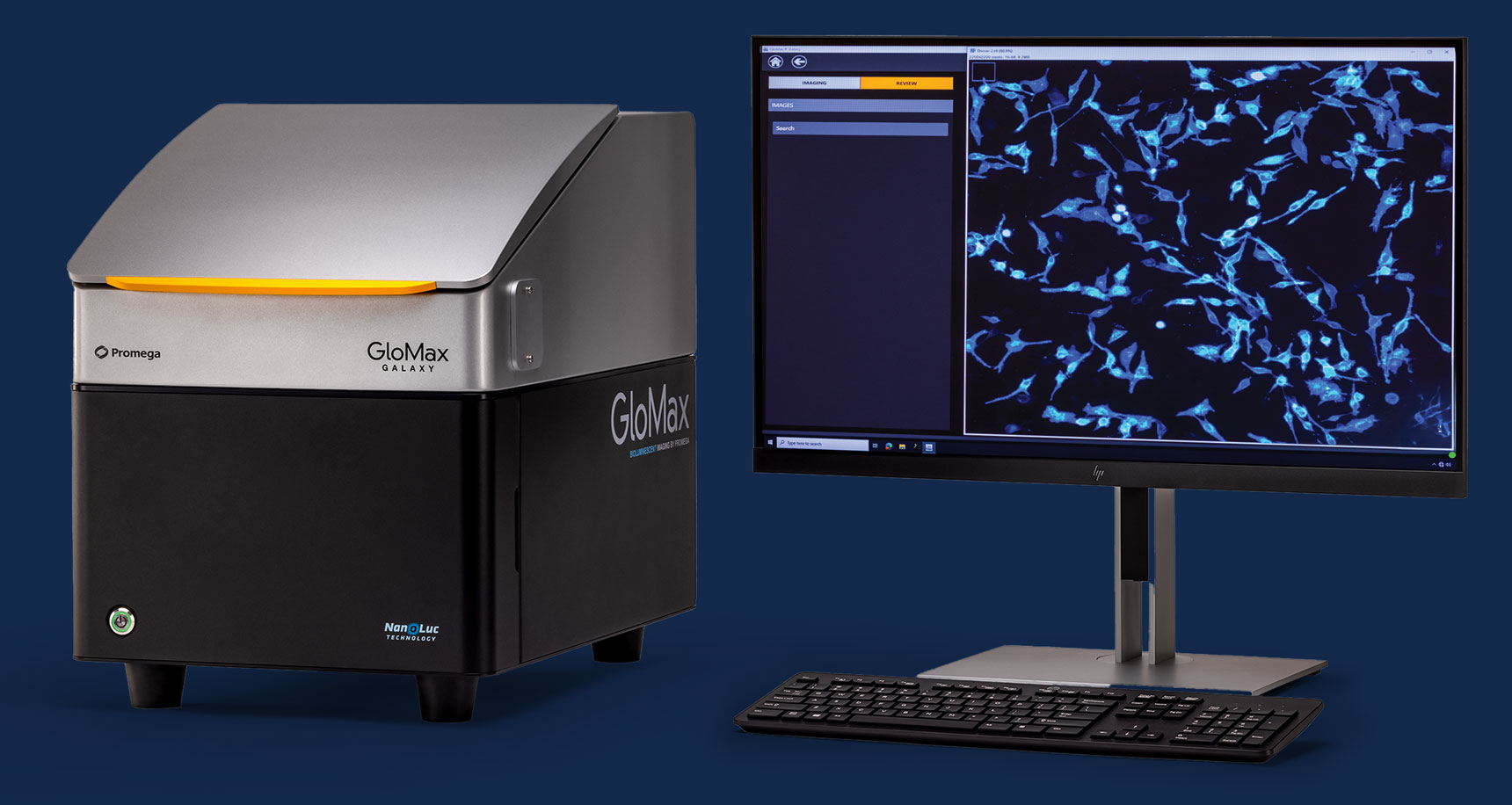
The GloMax® Galaxy Bioluminescence Imager combines the strength of bioluminescent and fluorescent reporters with live imaging capabilities. It is designed to enable imaging of our NanoLuc® Luciferase technologies (e.g., NanoBRET, NanoBiT, HiBiT and Lumit) to visualize various cellular processes:
- Protein:protein interactions
- Protein localization and translocation
- Protein degradation and stability
- Ligand:protein interactions (target engagement)
- Targeted cell killing

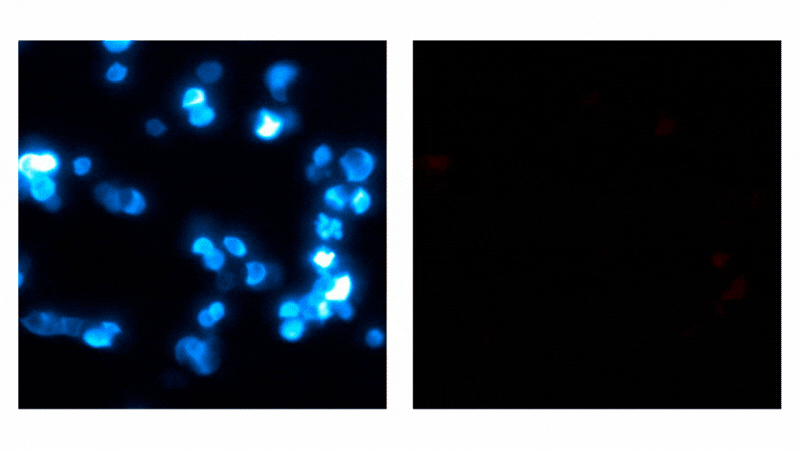
Detecting protein:small molecule interactions with NanoBRET® NanoGlo® Detection Systems. NanoBRET® Technology utilizes a bioluminescent donor and fluorescent acceptor to visualize complex protein:protein interactions, such as target engagement. HCT116 cells expressing a PRMT5–NanoLuc® fusion were supplemented with a fluorescent small molecule tracer. Before tracer addition, luminescent signal indicates energy is present on the donor protein (left; 3-minute exposures for 15 minutes). Binding of fluorescent tracer results in energy transfer and fluorescent signal (right; 3-minute exposures for 60 minutes). Videos were captured on the GloMax® Galaxy Bioluminescence Imager.
Illuminating Functional Biology
Webinar: Imaging NanoLuc® Luciferase Technologies with the GloMax® Galaxy Bioluminescence Imager.
See how bioluminescent assays and the GloMax® Galaxy Imager compliment studies of target engagement, receptor internalization, localization, and protein degradation.
What is Bioluminescent Live Cell Imaging?
Bioluminescent imaging in live cells is a technique that uses the light emission generated by a naturally catalyzed chemical reaction in biological organisms to visualize cellular processes, such as subcellular protein localization, in real time. Bioluminescent live cell imaging allows direct visualization of protein dynamics in living cells without the need for repeated sample excitation. How does bioluminescence work?
To learn more about the advantages and applications of this technique, explore our Bioluminescence Imaging Page.
NanoLuc® Luciferase reporters are well-suited for use in bioluminescent imaging studies. The extreme brightness means that exposure times can be reduced to only a few seconds, compared to the minutes required for other luminescent reporter proteins. Its small size also makes it less likely to perturb the normal biology or functionality of the fusion partner. Our Nano-Glo® Extended Live Cell Substrates have increased signal stability, allowing extended kinetic analysis lasting several hours to days.
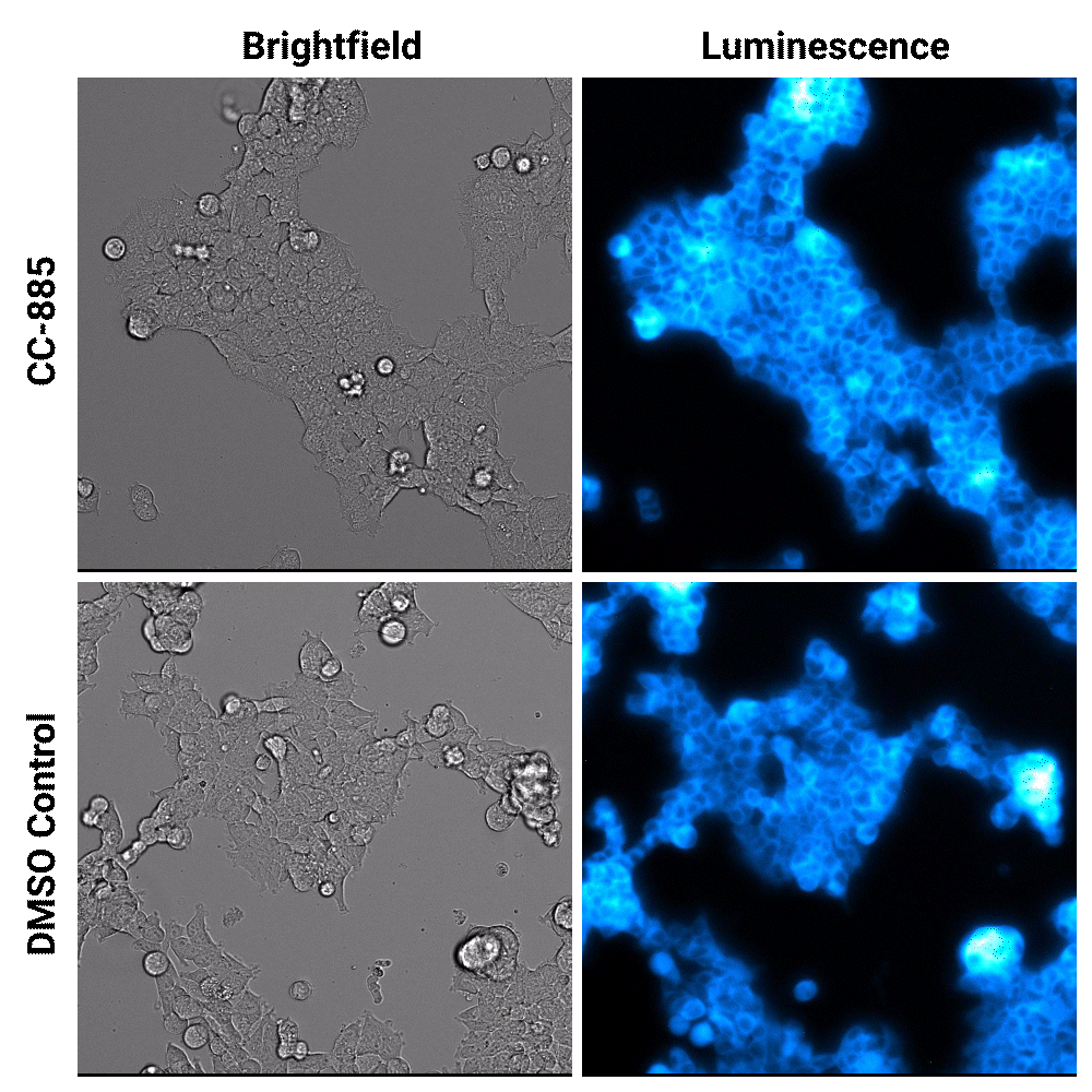
Targeted protein degradation over time. HEK293 cells expressing endogenous HiBiT-tagged GSPT1 and stably expressing LgBiT were treated with CC-885 degrader or DMSO control treatment. Assayed with Nano-Glo® Vivazine™ Live Cell Substrate and imaged over 5 hours using GloMax® Galaxy Bioluminescence Imager.
Dr. Li-Fang Chu's lab observed the exact timing of oscillating genes in early human development for the first time using bioluminescent live cell imaging. Read more in this blog

Featured Lab Manager Article: Unlocking Cellular Insights with a Bioluminescence Imaging System
This article outlines how bioluminescence imaging systems enhance experimental insight and highlights the critical features to evaluate when choosing the right platform for your research.
What is Fluorescent Live Cell Imaging?
Fluorescent live cell imaging is a technique that uses fluorescent proteins or dyes to visualize cellular structures and processes in real time. These fluorophores emit light when excited by specific wavelengths, allowing researchers to monitor events such as protein localization, organelle dynamics, and intracellular signaling. Learn more about fluorescence microscopy applications.
Fluorescent Ligands for Live Cell Imaging
We offer a large variety of fluorescent HaloTag® Ligands that enable live cell imaging. Explore our full suite of fluorescent ligands.
Fluorescent Live Cell Imaging Protocol
Interested in fluorescent live cell imaging, but not sure where to start? We provide comprehensive protocols for both rapid and no-wash live-cell imaging using our fluorescent HaloTag® Ligands. View the protocol to learn how to optimize a signal that is both robust and specific.
What is Live Cell Imaging of Spheroids and Organoids?
Spheroids and organoids are advanced 3D cell models that more closely mimic tissue structure and function than traditional 2D cultures. These models are widely used to study disease mechanisms, drug response and complex cellular interactions.
However, their structure also makes them more challenging to image. Live-cell imaging offers a powerful, non-invasive way to observe biological process over time, without damaging the model—making them ideal for long-term studies in 3D systems.
Want to learn more about 3D culture techniques? Explore our 3D Cell Culture Guide.
Monitor Cerebral Organoids Using HaloTag® Technologies
Explore how researchers are using HaloTag® and Janelia Fluor® dyes to illuminate neural networks within live, cerebral organoids. This white paper highlights pulse-chase imaging, inter-organoid communication, and long-term fluorescent labeling with low background, empowering detailed spatial analysis in 3D culture systems.
Visualize Organoids in Real Time
Webinar: Real-Time Non-Destructive Monitoring of Human Cerebral Organoids via NanoLuc®-HaloTag® Reporter System
See how real-time bioluminescence and fluorescence imaging with the NanoLuc®-HaloTag® system enables continuous, non-destructive tracking of neural differentiation and cerebral organoid development.
How Live Cell Imaging Is Used Across Research Applications
Cancer Research
Live cell imaging allows for the tracking of tumor cell behavior, including growth, invasion, and response to treatments. Cancer-specific markers and pathways can be monitored in real time using live cells.
Developmental Biology
Developmental processes like cellular proliferation, replication, and tissue formation are well-suited for study through live cell imaging. Understanding these processes requires visualizing the spatial and temporal patterns of gene expression and protein localization.
Immunology
Live cell imaging allows for the visualization of immune processes, including immune cell interactions, signaling pathways, and the immune response to pathogens. It can closely monitor inflammatory responses, such as gene activation in response to infection, and processes like phagocytosis.
Neuroscience
Live cell imaging enables the observation of neuronal activity, such as synaptic function and calcium signaling, within live neurons and larger neuronal networks. This technique allows for the study of neuron-specific genes and pathways that are implicated in neurodegenerative conditions over extended periods.
Microbiology
Microbial cells can be labeled with fluorescent or bioluminescent markers, enabling the study of host-pathogen interactions, biofilm formation, and microbial behavior. This approach is particularly useful for observing bacterial and viral infections, which is crucial for developing effective antimicrobial treatments.
Explore Bioluminescent Imaging Applications
See how researchers are using the GloMax® Galaxy Bioluminescence Imager to get more from their NanoLuc® Luciferase experiments. Explore proven imaging applications and find inspiration for your next discovery.
Additional Resources
Imaging
Learn about various imaging techniques, including live cell imaging, in vivo imaging, bioluminescence imaging and fluorescence microscopy.
In Vivo Imaging
Learn about in vivo imaging techniques including bioluminescence and fluorescence imaging, advantages, challenges, and applications in research.
Bioluminescence Imaging
Learn about bioluminescence imaging in whole animals and in live cells.