Lumit® Active IL-18 (Human) Immunoassay
No-Wash Immunoassay for Active IL-18
- Specific detection of mature/active IL-18; does not recognize pro-IL-18
- Fast add-and-read protocol
- Scalable to 96- or 384- well plates, protocols included
- Pack sizes show assay number for 96-well plates


Catalog Number:
Choose a product
Size
Catalog Number: W1910
Catalog Number: W1911
Catalog Number: W1912
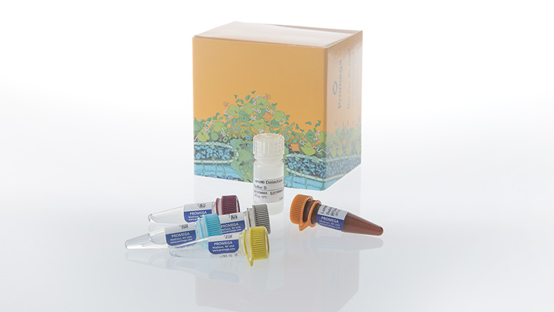
Catalog Number: W191A-C
Specific Detection of Active IL-18 Without Tedious Wash Steps
The Lumit® Active IL-18 Immunoassay quantitatively measures active IL-18 in cell culture samples using a simple, no-wash protocol. Just add labeled binding protein with treatment, add labeled antibody, add detection reagent and read luminescent signal using a standard plate-reading luminometer. The entire protocol is completed in less than 70 minutes! The assay can be used directly on cells in culture or culture medium samples transferred to a separate assay plate.
Note: The total number of assays per kit will vary based on final well volume.
For standard well volumes:
- 100 assay kits: applicable for 100 assays in 96-well plates or 400 assays in 384-well plates.
- 500 assay kits: applicable for 500 assays in 96-well plates or 2,000 assays in 384-well plates.
- 1,000 assay kits: applicable for 1,000 assays in 96-well plates or 4,000 assays in 384-well plates.
How the Lumit® IL-18 Immunoassay Works
Lumit® Flex technology is a luminescent structural complementation system composed of two small peptide tags (Peptide α and Peptide β) and a reagent-based polypeptide (Lumit® Flex Detection Protein). In the Lumit® Active IL-18 Immunoassay, a monoclonal anti–human IL-18 antibody and human IL-18 binding protein (IL-18BP) are labeled with Peptide α and Peptide β, respectively. In the presence of active IL-18, the two peptides are brought into proximity. When Lumit® Detection Reagent B (containing the Flex Detection Protein) is added, a functional luciferase is reconstituted and produces a bright luminescent signal in the presence of Lumit® Substrate. The signal is directly proportional to active IL-18 levels.
The assay specifically detects active IL-18 and does not recognize pro-IL-18 or IL-18 bound to endogenous IL-18BP.
For proper assay performance, hIL-18BP-β must be incubated with cells during treatment to capture active IL-18 immediately as it is released from cells. This is because active IL-18 is unstable in cell culture media. To see data demonstrating the instability of active IL-18, review this scientific poster.

Simple One-Plate Protocol Requires No Wash Steps

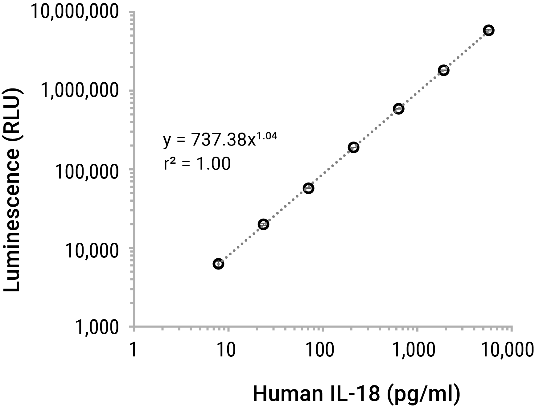
Broad Linear Range of Lumit® IL-18 Detection
The picomolar to nanomolar linear detection range of Lumit® IL-18 Immunoassay covers expected concentrations for most cell models.
|
Specification |
Lumit® IL-18 Immunoassay |
|
|
Dynamic Range |
7.8pg/ml–5,714pg/ml |
|
|
Limit of Detection (LOD) |
1.9pg/ml (3 SD above background) |
|
|
Minimal Detectable Dose (MDD) |
1.6pg/ml (2 SD above background) |
|
|
Assay Time |
70 minutes |
|
|
Sample Type |
Cell culture supernatants |
|
SD=Standard Deviation
Featured Webinar
Using Lumit® Technology to Address Inflammasome-Mediated Cytokine Release
In this webinar, Promega Sr. Research Scientist, Dr. Martha O’Brien, describes developing tools to measure inflammasome activation, including the Lumit® Active IL-18 Immunoassay and Lumit® IL-1β Immunoassay. Dr. O’Brien shows how Lumit® Assays can be multiplexed with other inflammasome assays to identify and rank the potency of inflammasome inhibitors.
Detect IL-18 Release Following Inflammasome Activation

The Lumit® Active IL-18 (Human) Immunoassay was used to quantitate secretion of active IL-18 from THP-1 cells following treatment. Active IL-18 is found in cell culture media samples following treatment with either nigericin or nigericin + LPS but not in untreated cells or cells treated with LPS alone. The amount of active IL-18 secreted was calculated using the standard curve. Data shown is an average of 3 replicates; error bars represent standard deviation.
Protocols
Specifications
Catalog Number:
Lieferumfang
| Produkt | Katalognummer | Größe |
|---|---|---|
Lumit® Detection Substrate B |
VB405A | 1 × 160μl |
Lumit® Detection Buffer B |
VB406A | 1 × 3.2ml |
Lumit® Flex Detection Protein |
VB720A | 1 × 450μl |
hIL-18BP-Peptide β, 1000X |
W192A | 1 × 20μl |
Anti-hIL-18 mAb-Peptide α, 1000X |
W193A | 1 × 20μl |
Lumit® Human Active IL-18 Standard |
W191A-C | 1 × 25μl |
SDS
Search for SDSAnalysezertifikat
Nutzungseinschränkung
For Research Use Only. Not for Use in Diagnostic Procedures.Lagerhinweise
U.S. Pat. No. 8,809,529, European Pat. No. 2635582, Japanese Pat. No. 5889910 and other patents and patents pending.
U.S. Pat. Nos. 9,797,889, 9,797,890, 10,107,800 and 11,493,504; European Pat. No. 2970412; Japanese Pat. Nos. 7280842 and 7532562; and other patents and patents pending.
Lieferumfang
| Produkt | Katalognummer | Größe |
|---|---|---|
Lumit® Detection Substrate B |
VB405B | 1 × 1.25ml |
Lumit® Detection Buffer B |
VB406B | 1 × 25ml |
Lumit® Flex Detection Protein |
VB720B | 1 × 4.5ml |
hIL-18BP-Peptide β, 1000X |
W192B | 1 × 200μl |
Anti-hIL-18 mAb-Peptide α, 1000X |
W193B | 1 × 200μl |
Lumit® Human Active IL-18 Standard |
W191A-C | 1 × 25μl |
SDS
Search for SDSAnalysezertifikat
Nutzungseinschränkung
For Research Use Only. Not for Use in Diagnostic Procedures.Lagerhinweise
U.S. Pat. No. 8,809,529, European Pat. No. 2635582, Japanese Pat. No. 5889910 and other patents and patents pending.
U.S. Pat. Nos. 9,797,889, 9,797,890, 10,107,800 and 11,493,504; European Pat. No. 2970412; Japanese Pat. Nos. 7280842 and 7532562; and other patents and patents pending.
Lieferumfang
| Produkt | Katalognummer | Größe |
|---|---|---|
Lumit® Detection Substrate B |
VB405A | 5 × 160μl |
Lumit® Detection Buffer B |
VB406A | 5 × 3.2ml |
Lumit® Flex Detection Protein |
VB720A | 5 × 450μl |
hIL-18BP-Peptide β, 1000X |
W192A | 5 × 20μl |
Anti-hIL-18 mAb-Peptide α, 1000X |
W193A | 5 × 20μl |
Lumit® Human Active IL-18 Standard |
W191A-C | 1 × 25μl |
SDS
Search for SDSAnalysezertifikat
Nutzungseinschränkung
For Research Use Only. Not for Use in Diagnostic Procedures.Lagerhinweise
U.S. Pat. No. 8,809,529, European Pat. No. 2635582, Japanese Pat. No. 5889910 and other patents and patents pending.
U.S. Pat. Nos. 9,797,889, 9,797,890, 10,107,800 and 11,493,504; European Pat. No. 2970412; Japanese Pat. Nos. 7280842 and 7532562; and other patents and patents pending.
Lieferumfang
| Produkt | Katalognummer | Größe |
|---|---|---|
Lumit® Human Active IL-18 Standard |
W191A-C | 1 × 25μl |
SDS
Search for SDSAnalysezertifikat
Nutzungseinschränkung
For Research Use Only. Not for Use in Diagnostic Procedures.Lagerhinweise
Resources
Fachartikel
- Lumit® Cytokine Assays: Interpolating Data with the GloMax® Discover
- Easier Detection of Inflammasome Activation
- High-Throughput Cytokine Detection with Lumit® Technology
- A Comprehensive Toolkit for NLRP3 Inflammasome Drug Discovery
- Immunoassay Guide
- White Paper: Lumit® Immunoassays: A Rapid and Sensitive Method for Analyte Detection
- Blog Article: Understanding Inflammation: A Faster, Easier Way to Detect Cytokines in Cells
Poster
- Poster: Lumit® Immunoassays: Bioluminescent, Sensitive, and Homogeneous Analyte Detection Using Labeled Antibodies
- Rapid and Sensitive Determination of Cytokine Release from Cells Without Sample Transfer
- A Lumit® Human IL-18 Assay Specifically Measures Active IL-18 Release in Cell Culture
- SLAS 2024 Lumit Cytokine Assay Automation for HTS
Zusätzliches Informationsmaterial
Related Products
Ähnliche Produkte
Lumit® IL-1β Human/Mouse Immunoassay
Quantifies inflammasome activation by measuring released IL-1β using a simple, no-wash protocol.
W6010, W6011, W6012, W7010, W7011, W7012, W116A-C, W119A-C
Lumit® IL-12 p70 (Human) Immunoassay
Quantitatively measures released IL-12 in cell culture samples using a simple, no-wash protocol that can be completed in 70 minutes or less.
W1850, W1851, W1852, W185A-C
Lumit® IFN-γ (Human) Immunoassay
Quantitatively measures released IFN-γ in cell culture samples using a simple, no-wash protocol.
W6040, W6042, W6041, W134A-C
Lumit® TNF-α (Human) Immunoassay
Quantitatively measures released TNF-α in cell culture samples using a simple, no-wash protocol.
W6050, W6052, W6051, W137A-C
Wird oft zusammen gekauft
Caspase-Glo® 1 Inflammasome Assay
A homogeneous, bioluminescent method to selectively measure the activity of caspase-1, an essential component of the inflammasome.
G9951, G9952, G9953
Thaw-and-Use Primary Effector Cells
Consistent and robust method for target cell killing.
CS3055C02, CS3055C03, CS3055B03, CS3055B06, CS3055A15, CS3055A18
CellTox™ Green Cytotoxicity Assay
Measures changes in membrane integrity. Kinetically monitors cytotoxicity up to 72 hours with multiplex capability.
G8741, G8742, G8743, G8731
GloMax® Discover System
High-performance microplate reader for detecting luminescence, fluorescence and absorbance.
GM3000