OncoMate® MSI Dx Analysis System
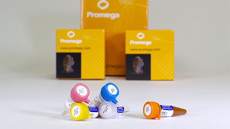
- CE-marked and available in select European countries
- Determine MSI status with Gold Standard microsatellite markers
- Functional test where detecting MSI demonstrates a deficiency in tumor DNA mismatch repair

Catalog Number:
Size
Catalog Number: MD3140
Accurately Determine MSI Status of Tumors Using the Gold Standard Method
The OncoMate® MSI Dx Analysis System is a fluorescent, multiplex PCR-based test to detect microsatellite instability (MSI). MSI is a form of genomic instability caused by the insertion or deletion of repeating bases within microsatellites during DNA replication due to the failure of the mismatch repair system (MMR) to correct these errors.
The OncoMate® MSI Dx Analysis System (Cat.# MD3140) is available in the following countries: Austria, Belgium, Czech Republic, Denmark, France, Germany, Hungary, Ireland, Italy, Luxembourg, Netherlands, Norway, Poland, Romania, Slovakia, Spain, Sweden, Switzerland and the United Kingdom. For information about availability in other regions, including the United States (FDA-approved) and China (approved for companion diagnostic use), please contact us.

MSI Testing Empowers Precision Medicine Through Actionable Insight
Uncover Hereditary Risk and Guide Confident Diagnostic and Management Decisions in Colorectal Cancer
Testing colorectal cancers for microsatellite instability (MSI) is critical for identifying patients with Lynch syndrome, enabling early detection of new or recurrent cancers in patients and their families. MSI-H status indicates defects in the DNA mismatch repair (MMR) system, In Lynch syndrome, that defect arises from an inherited pathogenic variant in an MMR gene, transmitted in an autosomal dominant pattern (1). PCR-based MSI testing provides direct evidence of MMR deficiency, helping clinicians assess hereditary cancer risk and guide appropriate management.
European clinical guidance consistently recommends that all colorectal cancers undergo universal tumor testing to identify probable Lynch syndrome, regardless of patient age or disease stage. This recommendation is reflected in ESMO clinical practice guidelines for localized and metastatic colorectal cancer (2–4), the EHTG/ESCP Mallorca guidelines (5), NICE diagnostics guidance DG27 (6) and the updated German S3 guideline (7). Universal testing triages large patient populations, identifying the smaller subset of patients who proceed to confirmatory germline sequencing.
The OncoMate® MSI Dx Analysis System is built for this purpose. The standardized, validated PCR-based workflow is scalable and cost-efficient for high-volume screening, requires as little as 1ng of input DNA and delivers results in 10 hours to support timely reporting.
Performance and Methodology to Meet Clinical Demands
Only Two Sections Required

FFPE sections with a tissue volume of 0.1mm3 to 2.0mm3. Tumor sample should have >20% viable tumor cells.
Minimal DNA Required

Uses only 1ng of amplification-quality DNA per reaction.
Assay Time Under 3 Hours

Quick, multiplexed reaction, balanced for accuracy and efficiency, goes from DNA to answer in as little as 2.5 hours.
Sensitive and Specific Results

Reliable MSI determination with strong agreement with IHC.
In a retrospective study of colorectal cancers, the OncoMate® MSI Dx Analysis System showed concordance with sequencing and immunohistochemistry (8) and is intended to identify patients that may benefit from further diagnostic testing.
Test Design and Marker Information
The OncoMate® MSI Dx Analysis System targets five mononucleotide repeat markers (BAT-25, BAT-26, NR-21, NR-24 and MONO-27) that were selected for high sensitivity and specificity to alterations in repeat lengths in samples containing mismatch repair defects (9,10). For quality control and sample authentication of matched tumor and normal samples, the system uses two pentanucleotide repeat markers (Penta C and Penta D) that were selected for their high level of polymorphism and lower degree of MSI.
Biomarker and Loci Information
Target Markers
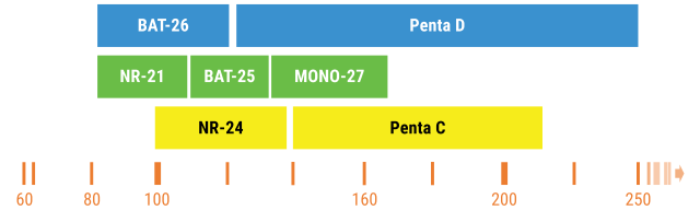
NCI recommended, Revised Bethesda Panel and two pentanucleotide markers (10).
Example Data
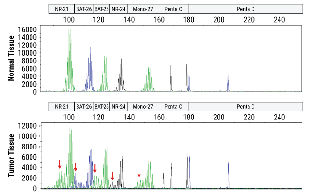
Instability is determined by fragment size analysis on a capillary electrophoresis instrument following PCR amplification of DNA from a patient's normal and tumor tissue samples. Unstable loci in the tumor tissue are indicated by red arrows.
Tumor to Answer Overnight
The OncoMate® MSI Dx Analysis System is part of a broader workflow that includes DNA extraction from FFPE tissue samples, quantitation of DNA, amplification of specific microsatellite markers using multiplex PCR, fragment separation by capillary electrophoresis, and data analysis and interpretation.

Isolate DNA
from FFPE samples

Quantitate DNA
using fluorescent DNA quantitation reagents and instruments

Amplify DNA
using the OncoMate® MSI Dx Analysis System

Calibrate the Dye Spectrum
using the OncoMate® 5C Matrix Standards

Separate and Detect Fragments
using a capillary electrophoresis instrument capable of fluorescent detection

Data Analysis and Reporting
using fragment analysis software

Turnaround Time:
In as little as 10 hours or overnight
Overview of the OncoMate® MSI Dx Analysis System Workflow

Compatibility Specifications
Specimens
Normal and tumor FFPE tissue samples with a volume of 0.1–2.0mm3. Tumor samples must contain at least 20% viable tumor cells.
Capillary Electrophoresis
Use with capillary electrophoresis instruments capable of:
- Fragment resolution of 1bp from 60bp to ≥300bp
- Sizing precision ≤0.15bp across a range from 60bp to ≥300bp
- Calibration and detection of 5 channel collection with excitation wavelengths from 480nm to 520nm and detection optics to capture from approx 500nm to 630nm
Fragment Analysis Marker Panels and Settings
Capillary electrophoresis data requires further analysis to assign marker and size information. We have provided fragment analysis marker panels and analysis settings for download. The provided settings were used during the performance evaluation of the OncoMate® MSI Dx Analysis System and may require optimization for your chosen capillary electrophoresis equipment.
Intended Use Statement: The OncoMate® MSI Dx Analysis System is a nonautomated qualitative PCR-based fragment-sizing test used to determine microsatellite instability (MSI) status in DNA purified from human formalin-fixed paraffin-embedded (FFPE) tissue samples derived from solid tumors of patients already diagnosed with colorectal cancer. The OncoMate® MSI Dx Analysis System is used to aid in diagnosing patients with Lynch syndrome, a hereditary cancer syndrome, by identifying colorectal cancer patients with MSI-High tumors who may benefit from additional germline testing. The OncoMate® MSI Dx Analysis System is not intended for use as an aid to drug selection for cancer patients.
The OncoMate® MSI Dx Analysis System generates allelic profiles from tumor and normal (non-tumor) FFPE tissue samples from the same patient using a multiplexed polymerase chain reaction (PCR) amplification of 5 mononucleotide DNA microsatellite markers (BAT-25, BAT-26, NR-21, NR-24 and MONO-27), followed by size separation of the amplified markers using fluorescence detection on a capillary electrophoresis instrument. MSI status is determined by comparing the allelic profiles. An alteration in the length of repetitive DNA sequences in the tumor cell DNA when compared to the normal cell DNA from the same patient indicates MSI. Normal and tumor tissue from the same patient must be tested at the same time, and data from both samples must be available for comparison for results to be valid.
Test results obtained using the product must be interpreted by healthcare professionals in conjunction with other clinical findings, family history and laboratory data. This product is intended for professional use only.
References
- Boland, C.R. and Goel, A. (2010) Microsatellite instability in colorectal cancer. Gastroenterology. 138(6), 2073–87.e3.
- Stjepanovic, N. et al. (2019) Hereditary gastrointestinal cancers: ESMO Clinical Practice Guidelines for diagnosis, treatment and follow-up. Ann. Oncol. 30, 1558–71.
- Argilés, G. et al. (2020) Localised colon cancer: ESMO Clinical Practice Guidelines for diagnosis, treatment and follow-up. Ann Oncol. 31(10), 1291–1305.
- Cremolini, C. et al. (2026) Metastatic colorectal cancer: ESMO Clinical Practice Guideline for diagnosis, treatment and follow-up. Ann Oncol. 37(6), 759–76.
- Seppälä, T.T. et al. (2021) European guidelines from the EHTG and ESCP for Lynch syndrome: an updated third edition of the Mallorca guidelines based on gene and gender. Br J Surg. 108(5), 484–98.
- National Institute for Health and Care Excellence. Molecular testing strategies for Lynch syndrome in people with colorectal cancer. HealthTech guidance HTG430 (formerly DG27). Published February 22, 2017. Accessed September 21, 2026. https://www.nice.org.uk/guidance/htg430.
- Vangala, D.B., Seufferlein, T. and Hüneburg, R. (2026) Lynch syndrome in context of the updated German S3 guideline colorectal cancer: implementation of universal MMR/MSI testing. Oncol Res Treat. 49(3), 165–173.
- OncoMate® MSI Dx Analysis System Technical Manual #TM595, Rev1, Promega Corporation.
- Bacher, J. et al. (2004) Development of a fluorescent multiplex assay for detection of MSI-High tumors. Dis. Markers 20, 237.
- Umar, A. et al. (2004) Revised Bethesda Guidelines for hereditary nonpolyposis colorectal cancer (Lynch syndrome) and microsatellite instability. J. Natl. Cancer Inst. 96, 261–8.
Seamless Support for Adopting OncoMate® MSI Dx in Your Lab
Our team is dedicated to partnering with you to ensure success and has broad experience training laboratories. We offer an online user training course as well as virtual and in-person training to get your lab up and running with the OncoMate® MSI Dx Analysis System more efficiently.

Medical Information
If you need assistance or access to medical information and resources, our Medical Affairs scientists are ready to help.
Protocols
Specifications
Catalog Number:
Lieferumfang
| Produkt | Katalognummer | Größe | Konzentration |
|---|---|---|---|
Water, Amplification Grade |
MD193A | 1 × 1,250μl | |
OncoMate® MSI 5X Master Mix |
MD280A | 1 × 200μl | |
Size Standard 500 |
MD500A | 1 × 100μl | |
OncoMate® MSI 5X Primer Mix |
MD705A | 1 × 200μl | |
2800M Control DNA |
MD810A | 1 × 25μl | 10ng/μl |
SDS
Search for SDSAnalysezertifikat
Nutzungseinschränkung
For In Vitro Diagnostic Use. This product is only available in certain countries.Lagerhinweise

Resources
Related Products
Wird oft zusammen gekauft
OncoMate® 5C Matrix Standard
Used to calibrate capillary electrophoresis instruments prior to running the OncoMate® MSI Dx Analysis System.
MD3850
Maxwell® CSC Instrument
Benchtop instrument for automated nucleic acid purification from a range of clinical sample types. For In Vitro Diagnostic Use.
AS6000
Maxwell® CSC DNA FFPE Kit
Automated DNA purification of DNA from FFPE samples for in vitro Diagnostic assays.
AS1350
Maxwell® CSC Blood DNA Kit
Automated genomic DNA isolation from blood for in vitro diagnostic Assays.
AS1321